Noncancerous Chest CT Performed Before Stage 1 Lung SBRT Predicts Survival
 Performing chest CT to detect noncancerous imaging markers before stereotactic body radiation therapy (SBRT) in stage 1 lung cancer patients improves survival prediction, compared with clinical features alone.
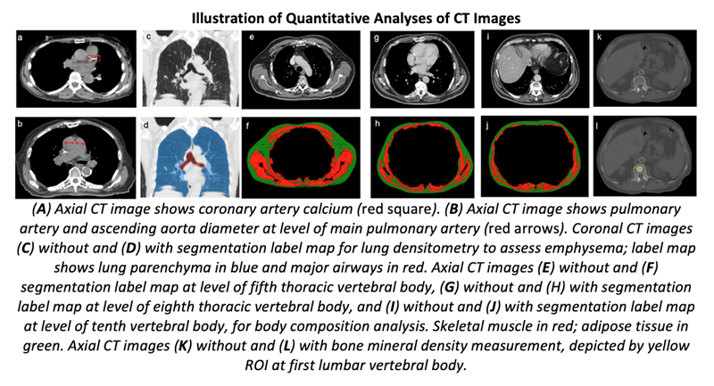
The retrospective study, published in the American Journal of Roentgenology, included 282 patients (168 female, 114 male; median age, 75 years) with stage I lung cancer treated with SBRT between January 2009 and June 2017. To quantify coronary artery calcium (CAC) score and pulmonary artery (PA)-to-aorta ratio, as well as emphysema and body composition, pretreatment chest CT was used. Associations of clinical and imaging features with overall were quantified using a multivariable Cox proportional hazards (PH) model.
“In patients undergoing SBRT for stage I lung cancer,” explained corresponding author and 2019 ARRS Scholar Florian J. Fintelmann, MD, “higher coronary artery calcium score, higher pulmonary artery-to-aorta ratio, and lower thoracic skeletal muscle index independently predicted worse overall survival.”
For stage I lung cancer patients treated with SBRT, CAC score, PA-to-aorta ratio, and skeletal muscle index showed significant independent associations with overall survival. (p<.05). The model including clinical and imaging features demonstrated better discriminatory ability for 5-year overall survival than the model including clinical features alone (AUC 0.75 vs. 0.61, p<.01).
“The PA-to-aorta ratio, which is readily quantifiable with electronic calipers during routine image review, was the most important predictor of overall survival,” the authors concluded.