Quantitative Micro-Elastography Breast Cancer Imaging System Receives FDA Breakthrough Device Status
 OncoRes Medical has received a Breakthrough Device designation from the US Food and Drug Administration to expedite development of its Quantitative Micro-Elastography (QME) Imaging System. The company's technology is designed to provide real-time tumor assessment that helps surgeons more accurately identify and remove cancerous tissue – an approach that could substantially improve outcomes in breast-conserving surgery and reduce repeat operations for women with breast cancer.
The company is currently raising a Series B round to fund ongoing product development and in vivo studies of its system. Equity funding to date has come from Australia's Medical Research Commercialization Fund, which is managed by Brandon Capital Partners, the country's leading life sciences venture capital fund manager. Additional funding has come from competitive grants and tax incentives from the Australian government.
While OncoRes Medical believes it can provide the biggest initial medical benefits with a focus on breast cancer, it is only the first of many clinical indications the company plans to pursue. QME is a platform technology that could eventually be applied to other solid cancers where complete tumor clearance is the foundation of curative treatment.
The QME Imaging System was originally designed to address the high rate of re-excision surgeries in breast cancer treatment, a well-recognized clinical issue known as "the other breast cancer epidemic." In the U.S., re-excisions occur in up to 35 percent of cases after initial breast conserving surgery. Repeat surgeries can lead to additional anxiety for patients as well as higher complication rates, poor cosmetic outcomes, and delayed treatments. There is also a significant economic burden, as re-excisions increase the total cost of breast cancer care in the U.S. by nearly $700 million each year.
"The Breakthrough Device designation highlights the potential of our QME technology to improve and even save lives by addressing a significant clinical issue," says medical doctor and OncoRes Medical CEO Katharine Giles, MBBS, MBA. "It will enable us to pursue a more efficient regulatory and reimbursement pathway in the U.S., ensuring that surgeons and patients have access to the clinical and potential economic benefits of this technology as soon as possible."
The FDA's Breakthrough Device program supports the timely development of technologies that have the potential to provide more effective treatment of life-threatening or irreversibly debilitating diseases or conditions. Under this program, OncoRes Medical will benefit from an expedited regulatory review process. The designation also has the potential to provide automatic reimbursement coverage for the QME Imaging System upon FDA approval.
The Problem
Each year approximately 331,000 U.S. women are diagnosed with breast cancer—a number that is expected to increase to more than 440,000 by 2030. To avoid a mastectomy, more than half of these women will opt for BCS to remove the tumor and preserve the appearance of their breast. Unfortunately, follow up surgeries are often required to ensure clear margins, or complete removal of the cancer.
There are no currently available technologies that can assist the surgeon to identify microscale residual tumor inside the surgical cavity, with surgeons still relying on their senses of sight and touch. As a result, incomplete tumor removal is only discovered three to seven days after surgery when the excised specimen is examined by a pathologist. Left untreated, positive margins on specimen pathology leaves patients at a twofold increased risk of breast cancer recurrence.
The Solution
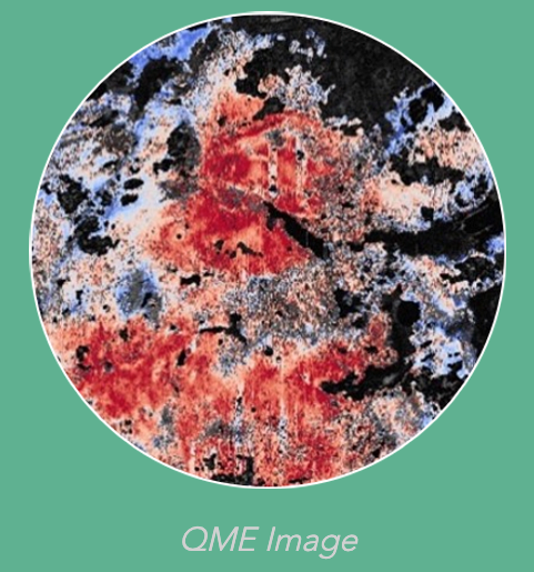
The OncoRes Medical QME Imaging System ensures complete tumor removal by facilitating the identification of residual cancer within the cavity during the initial surgery. It is well known that cancerous tissue varies in stiffness from healthy tissue, and the system combines optical coherence tomography (OCT) and micro-elastography technologies to help surgeons identify these variations in real-time. Similar to ultrasound, OCT uses light waves to produce images that are microscale in resolution, while elastography provides a quantitative measure of variation in tissue stiffness, or elasticity.
Following the excision of the main specimen during BCS, the handheld QME probe is applied to a region of interest within the breast cavity and a micro-scale three-dimensional map of the elastic properties of the scanned region is generated. These micro-scale maps of tissue stiffness offer surgeons an optical imaging method for assessing breast tissue for the presence of microscopic or otherwise non-palpable cancerous tissue remaining inside the breast cavity.
"The ability to detect residual cancer in the cavity at the time of surgery will be game-changing for both breast cancer patients and surgeons," says OncoRes Medical Chief Medical Officer Christobel Saunders, AO, MBBS, FRCS, FRACS, FAAHMS, an internationally recognized cancer surgeon and professor of surgical oncology at the University of Western Australia. "Surgeons will have greater control over the outcome of the procedure, while patients will have the peace of mind that comes from getting the first step of their breast cancer treatment right on the first try."
The Technology
A recent study of the QME Imaging System demonstrated the high accuracy of the technology for detecting cancerous tissue within 1 mm of the margin in tumor specimens excised during BCS. Applying an artificial intelligence algorithm further enhanced the accuracy of the readings, resulting in 100.0 percent sensitivity and 97.7 percent specificity. With confirmation of the technology's diagnostic accuracy, OncoRes Medical is currently planning trials in the U.S. and Australia to study its intended in-cavity clinical use.
There are currently no intraoperative tools that enable real-time, in-cavity imaging during BCS. Compared to other technologies on the market, the QME Imaging System also offers significant workow advantages as it images inside the surgical cavity and does not require contrast agents, ionizing radiation, or additional pre- or post-surgery procedures. In 2019, OncoRes Medical became the first Australian company to be accepted into the prestigious U.S.-based MedTech Innovator Accelerator Program, and was selected as that year's Value Award winner for its powerful value proposition to significantly improve the lives of patients with breast cancer.
