Robotic Bronchoscopy Helps Diagnose Lung Cancer
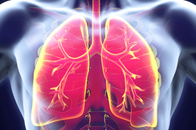 Screening those who are at high risk for lung cancer (people over age 50 who smoked one pack of cigarettes per day for 20 years or half a pack per day for 40 years) with an annual low-dose computed tomography (CT) scan has helped detect lung cancer in its earlier stages. If the CT scan finds an abnormality in a patient’s lungs, a bronchoscopy is generally recommended. During this common procedure, a doctor guides a thin tube equipped with a light and camera (bronchoscope) through the patient’s mouth or nose and into the lungs to obtain a tissue sample.
But there have been advances. Today, Yale Medicine physicians use a new form of technology called “robotic bronchoscopy,” which allows them to better reach smaller parts of the lungs. During a robotic bronchoscopy, the doctor uses a controller at a console to operate a robotic arm, which then guides the bronchoscope’s thin, flexible tube through the airways.
“With the regular bronchoscope, we can reach 20 to 30% of the airways, but with the robotic bronchoscope, we can reach 95% or more,” says Christopher Morton, MD, a Yale Medicine interventional pulmonologist. “The precision of the robot allows the tube to navigate tight turns and hard-to-reach areas of the lungs in order to obtain a biopsy of the lung tissue.”
But neither type of bronchoscopy is able to collect a sample from lymph nodes around the lungs and airways, which are important places to check for cancer and to determine if it has spread.
For that, doctors will perform an endobronchial ultrasound (EBUS) immediately after the bronchoscopy. The EBUS scope is similar to the bronchoscope, except that it has an ultrasound at its tip, which lets doctors see lymph nodes and guide a small needle to obtain a sample.
“The advantage of doing both the robotic bronchoscopy and the EBUS at the same time is that we get the most amount of information to accurately diagnose and stage the lung cancer with a low risk, and it is all done within one procedure,” explains Erin DeBiasi, MD, a Yale Medicine interventional pulmonologist.
With this information, doctors can quickly develop and implement any necessary treatment plan.